Of the X-ray apparatus the area to be X-rayed and the type of image receptor used the sensor or the phosphor plate these were all carefully considered. The patient is seated upright in the dental chair and should remove any removable dental appliances glasses or jewelry that could interfere with the X-ray beam.

Periapical Radiography Pocket Dentistry
To take a periapical exposure the hygienist or x-ray technician places a small photosensitive imaging plate coated with phosphorus into a sterile wrapper and inserts it into the patients mouth just like a conventional X-ray film card.

. You will need to bite firmly onto the device to keep it in place and provide a clear. Periapical film is held parallel to the long axis of the tooth using film-holding instruments. The film is placed parallel to the long axis of the tooth in question and the central x-ray beam should be directed perpendicular to the long axis of the tooth.
Disadvantages to the bisecting technique. Assessment of root morphology. RADIOGRAPHS Periapical Bitewing Occlusal.
Different techniques intra-oral bisecting angle intra-oral parallel and intra-oral parallel with bite registration. Ensure they are seated high enough so it is easy to see the occlusal. Each periapical x-ray shows a small section of your upper or lower teeth.
Parallel technique The image receptor is placed in a holder and placed in the mouth parallel to the longitudinal axis of the tooth under. The film is placed parallel to the long axis of the tooth to be radiographed and the central beam of X-ray is directed at right angle to the film and the teeth. The bisecting short-cone and paralleling long-cone techniques are two of the most commonly used techniques.
The bisecting technique may have to be used for patients unable to accommodate the film positioningdevice used in the paralleling technique. Film will be placed near your mouth using a metal rod with a ring attached to it. The sensor was placed on the.
Occlusal X-rays show full tooth development and placement 9. A long cone is used to take x-rays with paralleling exposure techniques. Assessment of root formation n completion.
Periapical x-rays are one of the most recommended and used x-rays in the field of dentistry. Changes to the angulation of the X-ray beam in relation to the teeth and film can help diagnosis and treatment by producing images which provide additional information not alway. Different techniques and instruments are used to drain and decompress large periapical lesions ranging from placing a stainless steel tube into the root canal exhibiting persistent apical exudation 202 204 which is non-surgical decompression to placing polyvinyl or polyethylene tubes through the alveolar mucosa covering the apical lesion which is surgical.
Extraoral radiograph Panoramic X-ray Tomograms Cephalometric projections Sialography Computed tomography 10. Periapical Lesion Diagnosis Support System Based on X-ray Images Using Machine Learning Technique. Since the slope and curvature of the dental arches and the alveolar processes will not permit the film to be held close to the teeth.
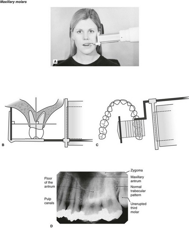
Bisecting angle and paralleling. The patient was positioned upright with hisher mouth was opened as wide as possible to allow the X-ray beam to pass to the sensor unobstructed from the opposite side of the mouth. By using a filmsensor holder with fixed image receptor and.
The Bisecting Angle Technique is an alternative to the paralleling technique for taking periapical films. The X-ray is taken and the exposed plate is then loaded into a scanner or processor which reads the image. The paralleling technique is recommended for routine periapical radiography but there are some instances when it is very difficult due to patient anatomy or lack of cooperation.
The X-ray apparatus that was used to obtain the periapical radiographs was the Sommo Gnatus Ribeirão Preto SP Brazil with an electric regime of 70kVp 7mA and with a. The X-ray tubehead is then aimed at right angles vertically and horizontally to both the tooth and the image. They are the basics in the field of dental radiographs and are used to a great extent in several branches of dentistry in order to ensure that the procedures are done well and the teeth are not harmed in any form.
Periapical radiographic techniques during endodontic diagnosis and treatment Int Endod J. Find the long axis of the tooth and then find the long axis of the image receptor as it is placed next to the tooth. The extraoral periapical radiographic technique was performed for both maxillary and mandibular teeth using Newman and Friedman technique2.
With this technique the film is placed parallel to the long axis of a tooth allowing the X-ray to be focused perpendicular to the long axis of the tooth. Periapical X-rays. The central ray is directed to pass at a perpendicular angle to both the tooth and the film.
For this purpose a special technique of periapical radiography was developed by Gordon M. In these situations the bisecting angle technique may be used. Implant site assessment and.
Both techniques have advantages and disadvantages. The central rays of the x-ray beam could either be directed perpendicular to the long axes of the teeth Or perpendicularly to the plane of the image receptor but not perpendicular to both at the same time. Paralleling Technique for Periapical X-rays The paralleling technique results in good quality x-rays with a minimum of distortion and is the most reliable technique for taking periapical x-rays.
Bite registration is of great importance for taking reproducible periapical x- rays in studying the dynamic processes in apical periodontal zone. By using a film sensor holder with still. The image receptor is placed in a holder and positioned in the mouth parallel to the long axis of the tooth under.
Assessment of relationship of roots to various vital structures. 1 Department of Pediatric Dentistry School of Odonto-stomatology Hanoi Medical University Dong Da Hanoi Vietnam 26 Department of. Periapical radiographs provide important information about the teeth and surrounding bone.
Vo TN Ngoc 1 Do H Viet 2 Le K Anh 3 Dinh Q Minh 4 Le L Nghia 5 Hoang K Loan 6 Tran M Tuan 7 Tran T Ngan 8 Nguyen T Tra 9. There are two types of techniques used for periapical radiographs. Periapical X-rays are used to detect any abnormalities of the root structure and surrounding bone structure.
Periapical views are used to record the crowns roots and surrounding bone. These patients may include adults with low palatal vaults and children. Periapical radiography is a commonly used intraoral imaging technique in radiology and may be a component of your radiologic examination.
Fitzgerald called as paralleling or long cone technique. Single periapical radiographs are often made of individual teeth or groups of teeth to obtain information for treatment or diagnosis of localized diseases or abnormalities. How periapical x-rays are taken.
The X-ray head is directed at right angles vertically and horizontally of both the tooth and the image receptor. These x-rays are often used to detect any unusual changes in the root and surrounding bone structures.

Periapical Radiography Pocket Dentistry

Periapical Radiography Pocket Dentistry

Periapical Radiography Pocket Dentistry

How Make Periapical X Ray

Periapical Radiography Pocket Dentistry
7 Periapical Radiography Pocket Dentistry
Periapical Radiography Pocket Dentistry

Periapical Radiography Pocket Dentistry
0 comments
Post a Comment